


TEST YOUR KNOWLEDGE

A TODDLER WITH LIMPING
SH Keah FRACGP, AM, Family Physician, Elizabeth Medical Centre
Address for correspondence: Dr Keah Say Hien, Elizabeth Medical Centre Sdn Bhd, 1-14, Jalan Arab, 84000 Muar, Johor, Malaysia. Tel: 06-9535335, Fax: 06-9543100. Email: drkeah@hotmail.com
Keah SH. Test Your Knowledge: A toddler with limping. Malaysian Family Physician. 2007;2(2):89
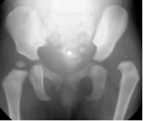
Sandy K, a 17 months old toddler, was brought by her mother for a medical examination in view of obtaining a life insurance. She is the only child. According to the mother, the child was noted to be limping since she started walking at 1 year of age. She brought her to see a traditional medicine sinseh for some massage to straighten her ‘tight muscles’. X-ray of her hips is shown in the figure below.
Questions
- What is the diagnosis?
- What physical findings can you elicit?
- What are the risk factors for this condition?
- What are the screening recommendations for this condition?
- What diagnostic tests are available?
- What treatment options can be offered in this case?
Answers
- Developmental dysplasia of the left hip. The name development dysplasia of the hip (DDH) is preferred to the older term (congenital dislocation of the hip) as it is a developmental disorder with variable manifestations and not always detectable at birth.
- Trendelenberg gait, limp length discrepancy, extra thigh fold, limited abduction of affected hip. The Barlow and Ortolani tests are positive only in the first 2-3 months of life; hence it is not relevant in this case.
- Risk factors for DDH include breech, female, first born and a positive family history. Sandy was delivered by Caesarean section for breech presentation (note: she is also female and first born).
- Some professional society (e.g. American Academy of Pediatrics) recommended routine screening hip examinations in the newborn nursery and then at every well child visit until walking age. However, the most recent guideline from the US Preventive Services Task Force did not find enough evidence to justify routine newborn screening of this condition.
- Ultrasound and X-rays. In newborn less than 4-6 months of life, imaging of the hip joint is best achieved with ultrasound. Within this age range, the femoral head and acetabulum are primarily cartilaginous. The femoral head usually ossifies after 4-6 months of age, thus plain radiographs is the most valuable imaging modality at this stage. These diagnostic imaging procedures are primarily used to confirm an abnormal physical finding. However, in high risk cases, one should have a lower threshold in getting the appropriate investigations at the appropriate time even though the physical examination may be normal.
- (i) Conservative treatment. The natural history of untreated DDH is likely to be a painless functional range of motion of the hip in early adulthood followed by osteoarthritis of the hip at around the fifth decade. The symptomatic hip with degenerative arthritis would then require a total hip arthroplasty. (ii) Closed reduction under general anesthesia with a preceding period of skin traction may be attempted. (iii) Open reduction. Reduction (open or close) must be done with great care to avoid avascular necrosis of the femoral hip. This will involve some variety of rebuilding of the femoral angle and the acetabulum together with releasing of the adductor which is causing the contracture. An early referral for an orthopedic consult is thus required in this patient.
Further readings
- Staheli LT. Paediatric Orthopedic Secrets, 2nd edition. Hanley & Belfus. 2003; 281-6
- Storer SK, Skaggs DL. Developmental dysplasia of the Hip. Am Fam Physician. 2006;74(8):1310-6 [PubMed] [Full text]
- Committee on Quality Improvement, Subcommittee on Developmental Dysplasia of the Hip. Clinical Practice Guideline: Early detection of developmental dysplasia of the hip. Pediatrics. 2000;105(4Pt1):896-905 [PubMed] [Full text]
- US Preventive Services. Screening for developmental dysplasia of the hip: recommendation statement. Pediatrics. 2006; 117(3):898-902 [PubMed] [Full text]
- Teo ELHJ. Clinics in Diagnostic Imaging (69). Bilateral developmental dysplasia of the hip. Singapore Med J. 2002;43(1):49-52 [PubMed] [Full text]