

Welcome to the
Malaysian Family Physician, a peer-reviewed open-access journal of family practice
and primary care research.
CASE REPORT

ETHAMBUTOL OCULAR TOXICITY IN A PATIENT WITH PULMONARY TUBERCULOSIS
– A CASE REPORT
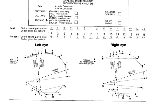
Figure 2. Farnsworth D-15 Hue Test at presentation showing bilateral asymmetrical tritanomaly.

Figure 3: Farnsworth D-15 Hue Test at 2 months after discontinuation of ethambutol showing normal color vision.
DISCUSSION
Ethambutol is one of the first-line drugs for tuberculosis and Mycobacterium avium complex (MAC) infections. Optic neuropathy is a well documented side-effect of ethambutol. The incidence of this reaction is proportional to the dose of ethambutol and is observed in 15% of patients receiving 50 mg/kg per day, in 5% of patients receiving 25 mg/kg per day, and in less than 1% of patients receiving daily doses of 15 mg/kg.1
Early-onset toxicity is an idiosyncratic reaction and is generally irreversible while delayed-onset toxicity is dose dependent due to zinc-chelation property of the drug and is generally reversible. Frequently, patients who developed optic neuropathy have no detectable risk factors.2
According to Kollner’s rule, retinal diseases cause blue-yellow colour vision defects whereas optic nerve diseases affect red-green discrimination.3 This distinction is however not reliable.4 Schneck et al5 demonstrated that in optic neuritis, blue-yellow defects tend to be slightly more common in the acute phase of the disease.
Polak et al6 reported that blue-yellow colour defects were the commonest and earliest defect in patients with ethambutol-induced optic neuropathy without any visual symptoms. Kumar et al noted that all the patients in their study had blue-yellow colour vision defects.2
Chest physicians, cardiologists, rheumatologists, urologists and internists routinely prescribe drugs that have the potential to cause ocular toxicity. Unfortunately, many non-ophthalmologists especially internists use ISP to screen for acquired colour defects. In most instances, it is the only modality available outside the ophthalmology department to test for colour vision abnormalities.
The ISP were designed specifically for the screening of congenital red-green colour deficiencies. Therefore, the use of ISP for the detection of acquired blue-yellow colour deficiency is not appropriate. Moreover, unless a baseline ISP test was done, the ISP is not able to differentiate congenital from acquired red-green defects.
The Farnsworth Panel D-15 Hue test, on the other hand, is a quick and convenient way to screen for colour vision deficiencies. The test can be done at the bedside. It is as easy to administer as the ISP test. It can differentiate congenital from acquired colour vision defects. The severity of colour defect was reflected by the number of crossing errors in the chart.7
For congenital colour defects, the patterns of the dichotomous analysis chart are very precise.4 In our patient, the dichotomous analysis patterns are irregular and asymmetry. This is characteristics for acquired colour defect, where the eyes were affected bilaterally but with varying severity.
Subtle blue-yellow defects can only be detected with the Lanthony Desaturated D-15 Test.8 However, there is considerable within-subject variability in test results. Lanthony Desaturated D-15 Test can be complicated by the wide variety of fine colour discrimination abilities in people with normal colour vision. Subtle loss of colour discrimination can also occur with aging.9
Ethambutol toxicity can occur even at the lowest recommended dosage levels.10 Vision loss can be severe and permanent.2 Early detection and immediate therapy discontinuation are the only effective management that can halt the progression of vision loss and allow recovery of vision.8
In our patient the ethambutol ocular toxicity was diagnosed by detecting blue-yellow colour defect using Farnsworth Panel D-15 hue test, and the drug was immediately discontinued. A repeat test after 2 months showed complete recovery of blue-yellow colour defect. Thus, this case report illustrates the importance of regular monitoring of patients receiving ethambutol for blue-yellow defect using the Farnsworth Panel D-15 hue Test.
REFERENCES
- Petri WA, Chapter 48: Antimicrobial Agents. In: Hardman JG, Limbrid LE, Gillman AG. Goodman and Gillman’s The Pharmacological Basis of Therapeutics, 10th edition, New York, McGraw-Hill, 2001, pg 1280.
- Kumar A, Sandramouli S, Verma L, et al. Ocular ethambutol toxicity: is it reversible? J Clin Neuro-ophthalmol. 1993;13(1):15-17 [PubMed]
- Vander JF, Gault JA. Ophthalmology Secrets, 2nd edition. Pennsylvania: Mosby Elsevier, 2002, pg 1.
- American Academy of Ophthalmology. Basic Science and Clinical Course. Section 12: Retina and Vitreous. San Francisco: American Academy of Ophthalmology, 2005, pg 44.
- Schneck ME, Hargerstrom-Portney G. Color vision defect type and spatial vision in the optic neuritis treatment trial. Invest Ophthalmol Vis Sci. 1997;38(11):2278-89 [PubMed] [Full text]
- Polak BC, Leys M, van Lith GH. Blue-yellow colour vision changes as early symptoms of ethambutol oculotoxicity. Ophthalmologica 1985;191(4):223-6 [PubMed]
- Good GW, Schepler A, Nichols JL. The reliability of the Lanthony Desaturated D-15 test. Optom Vis Sci 2005;82(12):1054-9 [PubMed]
- Chan RYC, Kwok AKH. Ocular toxicity of ethambutol. Hong Kong Med J. 2006;12(1):56-60 [PubMed] [Full text]
- Lakowski R. Age and color vision. Adv Sci 1958;15:231-6
- Melamud A, Kosmorsky G, Lee MS. Ocular ethambutol toxicity. Mayo Clin Proc. 2003;78(11):1409-11 [PubMed] [Full text]