

Welcome to the
Malaysian Family Physician, a peer-reviewed open-access journal of family practice
and primary care research.
TEST YOUR KNOWLEDGE

Respiratory Clinics
LEFT-SIDED HYDROPNEUMOTHORAX IN A YOUNG MALE: IMPORTANCE OF CLINICAL AND RADIOLOGICAL MARKERS IN ARRIVING AT AN AETIOLOGICAL DIAGNOSIS
R Khajotia, MBBS(Bom), MD(Bom), MD(Vienna), FAMA(Vienna), FAMS(Vienna)
Associate Professor in Internal Medicine and Pulmonology, International Medical University Clinical School,
Seremban, Negeri Sembilan, Malaysia. Email: rumi@imu.edu.my
ST Kew, MBBS(Sing), FRCP(Lond, Glasgow, Edin), FRACP(Hon), FRCPI
Professor and Head, Department of Internal Medicine, International Medical University Clinical School,
Seremban, Negeri Sembilan, Malaysia. Email: kew@imu.edu.my
YL Cham, MD(USM), MRCP(UK)
Medical Specialist, Hospital Tuanku Ja’afar, Seremban, Negeri Sembilan, Malaysia. Email: yeelingcham@gmail.com
Address of correspondence: Dr Rumi Khajotia, Associate Professor in Internal Medicine and Pulmonology,
International Medical University Clinical School, Jalan Rasah, 70300 Seremban, Negeri Sembilan, Malaysia
R Khajotia, ST Kew, YL Cham. Left-sided hydropneumothorax in a young male: Importance of clinical and radiological markers in arriving at an aetiological diagnosis. Malaysian Family Physician. 2009;4(1):41-43
CASE REPORT:
A 30-year old male patient presented to the Accident & Emergency (A&E) Department with a history of left-sided chest pain since 4 hours. Following a bout of coughing, he experienced a left-sided chest pain. The pain was diffusely present over the chest wall both anteriorly and posteriorly. There was no history of accompanying sweating, palpitation or vomiting. There was history of low-grade fever since one month which had now become high-grade since the past ten days. There was no other significant past medical history.
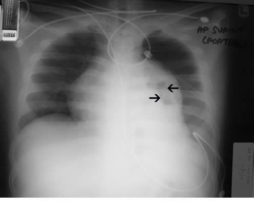
On general examination, the patient was tachypnoeic. His pulse rate was 106/min, regular and of good volume. His BP was 130/84 mmHg. On examination of the chest, the left hemithorax moved less than the right. Percussion revealed a hyperresonant note over the left upper and mid zones, both anteriorly and posteriorly, and a dull note over the left lower zone posteriorly. On auscultation, there was absence of breath sounds on the left side anteriorly. However, tubular bronchial breath sounds were heard in the left interscapular region posteriorly and vocal resonance was also increased in the same area. The right lung was normal. Chest x-ray showed a left-sided hydropneumothorax with complete collapse of the left lung (fig. 1). The mediastinum was shifted to the right side. The right lung appeared to be normal. Four hours after admission, an intercostal drainage tube was inserted on the left side in the 4th left intercostal space in mid-axillary line. About 500 ml of thick greenish-yellow fluid was drained.
Figure 1. A left-sided hydropneumothorax (white arrow) with complete collapse of the left lung (black arrows)

One week later, breath sounds continued to be significantly reduced on the left side with persistent bronchial breath sounds in the left interscapular region. The drainage column still continued to move on deep inspiration and bubbles were noted in the drainage tube on coughing. A chest radiograph was repeated (fig. 2). Based on the clinical and radiological findings, it was felt that a high-resolution CT scan (HRCT) of the chest needed to be done.
Figure 2. Semi-expanded left lung with areas of hyperluscency suggestive of lung parenchymal cavitation (black arrows).
QUESTIONS
- What do the clinical findings indicate?
- What do the serial chest radiographs (on admission and one week later) indicate, and why is an HRCT scan of the chest necessary in this patient?
- What are the findings on the HRCT chest scan?
- Taking into consideration the clinical and radiological findings, what is the likely aetiology of this patient’s condition?
- What other investigations would help to confirm the diagnosis?
- How would you explain the presence of the purulent pleural fluid?
Figure 3. High-resolution (HRCT) scan of the chest showing areas of cavitation in the semi-expanded left lung (gray arrows) and presence of a left-sided pleural effusion (gray arrow.

ANSWERS
- The acute presentation and clinical findings on admission are indicative of left hydropneumothorax and a collapsed left lung. However, the presence of bronchial breath sounds and increased vocal resonance in the left interscapular region also indicate the presence of an air-leak (broncho-pleural fistula) in the left lung. One week later, continued diminished breath sounds and presence of bronchial breath sounds are indicative of a persistent left lung collapse with an air leak. This is also corroborated by the fact that even one week later the air column continues to move on deep inspiration and air bubbles appear in the drainage tube on coughing. Hence, there is a strong clinical indication that a persistent air-leak is present. An alternative explanation could also be that this is a “trapped lung” which is failing to expand due to surrounding scarring and fibrous tissue. The bronchial breath sounds and increased vocal resonance in this case could be attributed to good sound conduction through a patent bronchus-to-collapsed lung-to-chest wall. Hence taking into consideration the important clinical parameters namely, presence of purulent pleural fluid and a persistent lung collapse (either due to a bronchopleural fistula or a trapped lung), a chronic infective pathology appears to be the most likely aetiology.
- The chest radiograph on admission shows a left-sided hydropneumothorax (presence of an air-fluid level) with a complete collapse of the left lung (Fig.1). One week later, following intercostal drainage the left lung has failed to significantly expand. However, the semi-expanded lung provides some valuable information. It shows the presence of a large area of hyperluscency indicative of areas of parenchymal cavitation(Fig. 2). The pleura also appears thickened. Taking into consideration the clinical and radiological findings it is felt that we are dealing with a chronic, infective, cavitary lung pathology. A persistent air-leak could have occurred due to rupture of a cavity. Since the left lung has failed to expand adequately and the semi-expanded lung is suspicious of underlying cavitations, an HRCT scan of the chest is felt necessary to confirm the findings.
- The HRCT scan of the chest confirms our clinical and radiological suspicions. It shows multiple cavities in the upper and lingular lobes of the left lung (Fig. 3). Surrounding areas of parenchymal consolidation are also seen. The pleura is also thickened with presence of air and fluid in the pleural cavity.
- Taking into consideration the clinical and radiological (including CT scan) findings, the likely aetiology is “cavitary pulmonary tuberculosis”.
- Once a chronic, infective pathology was suspected, the first likely thought was pulmonary tuberculosis. This was because the pathology appeared to be chronic and cavitary with presence of fluid in the pleural cavity. Other investigations done were sputum smear and culture examination for acid-fast bacilli (AFB), full blood count and erythrocyte sedimentation rate (ESR). The sputum was strongly positive for AFB (8+) on smear examination. The AFB culture report is awaited. The total leucocyte counts were elevated and the ESR was 80 mm/1hr.
- The presence of purulent pleural fluid can be explained by the fact that there was probably a long standing tuberculous pleural effusion in this patient which was not diagnosed. Consequently it developed bacterial superinfection and became purulent. Therefore, the presence of purulence does not rule out underlying tuberculous infection. The presence of significant pleural thickening (on HRCT scan) further substantiates the presence of long-standing pleural effusion in this patient. The initial low-grade fever was probably due to the underlying tuberculous infection and as the fluid developed bacterial superinfection the fever became high-grade.
In conclusion, it is apparent that clinical and radiological markers played a vital role in arriving at an aetiological diagnosis initially, which was further confirmed by presence of acid-fast bacilli (AFB) in the patient’s sputum.
REFERENCES:
- Palomino JC, Leao SC, Ritacco V. Tuberculosis 2007: From Basic Science to Patient Care. First Edition. www.tuberculosistextbook.com
- Crofton and Douglas’ Respiratory Diseases, Fifth Edition. Blackwell Science, 2008