

Welcome to the
Malaysian Family Physician, a peer-reviewed open-access journal of family practice
and primary care research.
REVIEW ARTICLE

THYROID ASSOCIATED OPHTHALMOPATHY – A REVIEW
PS Mallika1, MS; AK Tan1, MD; S Aziz2, MS; SAR Syed Alwi1, MMed; MS Chong2, MS; R Vanitha2 MS; G Intan2, MS.
1Department of Ophthalmology, Faculty of Medicine and Health Sciences, University Malaysia Sarawak, Kuching, Sarawak, Malaysia (Mallika Premsenthil, Tan Aik Kah, Syed Alwi Abdul Rahman)
2Department of Ophthalmology, Sarawak General Hospital, Kuching, Sarawak, Malaysia (Mohamad Aziz Salowi, Chong Min Siew, Vanitha Hema Ratnalingam , Intan Gudom)
Address for correspondence: Dr. Mallika Premsenthil, Lecturer, and Ophthalmology Unit, Faculty of Medicine and Health Sciences, University Malaysia Sarawak, Lot 77, Seksyen 22 Kuching Town Land District, Jalan Tun Ahmad Zaidi Adruce, 93150 Kuching, Sarawak, Malaysia. Tel +6082 416550 Fax + 6082 422564 Email: pmallika@fmhs.unimas.my
Conflict of interest: None
ABSTRACT
Thyroid associated ophthalmopathy is an autoimmune disorder affecting the orbital and periorbital tissues. Hyperthyroidism is commonly associated with thyroid associated ophthalmopathy, however in 5% to 10% of cases it is euthyroid. Genetic, environmental and endogenous factors play a role in the initiation of the thyroid ophthalmopathy. Smoking has been identified as the strongest risk factor for the development of the disorder. The pathogenesis involves activation of both humoral and cell mediated immunity with subsequent production of gycoaminoglycans, hyaluronic acid resulting in oedema formation, increase extraocular mass and adipogenesis in the orbit. The natural history of the disease progresses from active to inactive fibrotic stage over a period of years. Diagnosis is mainly clinical and almost all patients with ophthalmopathy exhibit some form of thyroid abnormality on further testing. Treatment is based on the clinical severity of the disease. Non-severe cases are managed by supportive measures to reduce the symptomatology and severe cases are treated by either medical or surgical decompression. Rehabilitative surgery is done for quiescent disease to reduce diplopia and improve cosmesis.
Key words: Thyroid eye disease, autoimmunity, smoking, corticosteroids, radiotherapy, surgical decompression, rehabilitative surgery.
Mallika PS, Tan AK, Aziz S, Syed Alwi SAR, Chong MS, Vanitha R, Intan G. Thyroid Associated Ophthalmopathy – A Review. Malaysian Family Physician. 2009;4(1):8-14
INTRODUCTION
Thyroid associated ophthalmopathy (TAO) is also known as, thyroid eye disease (TED), Graves’ ophthalmopathy/orbitopathy (GO), dysthyroid ophthalmopathy, thyrotoxic exophthalmos and other terms. It is an autoimmune process which affects the thyroid gland, orbital and periorbital tissue and uncommonly the pretibial skin or digits (thyroid acropachy). The individual components can occur together or separately. It is the most frequent extrathyroidal manifestation of Graves’ disease. Although TAO is often associated with hyperthyroidism, it may occur in primary hypothyroidism, Hashimoto’s thyroiditis, and sometimes in euthyroid individuals.1-3 The incidence and prevalence of Graves’ disease is 0.1% and 1% respectively. The clinical signs include widening of the palpebral fissure, eye lid retraction, lid lag, conjunctival congestion, chemosis, proptosis, corneal exposure, restrictive myopathy and optic neuropathy. In majority of cases the ocular manifestations are mild, and severe form of the disease affects 3% to 5% of individuals.4
METHODOLOGY
All our reference articles were obtained from Pubmed. The key words for search were thyroid ophthalmopathy, thyroid orbitopathy, thyroid associated ophthalmopathy, ocular manifestations of thyroid, ocular features of Graves’ disease, thyroid eye disease, and Graves’ ophthalmopathy etc. We used the MeSH database and journal database for our search and our search limits were articles in English and age above 1 year.
FREQUENCY
The exact incidence of ophthalmopathy is not clear. The prevalence of TAO (thyroid associated ophthalmopathy) in patients with GD (Graves’ disease) in Caucasian population is generally thought to be between 25% and 50 %.5,6 Bartley7 reported, in a population- based setting in USA, an annual incidence rate of 16 cases per 100,000 population per year for women, and 2.9 cases for men. In Malaysia, Lim et al8 reported a higher prevalence rate (34.7%) of thyroid associated ophthalmopathy in three populations of Asian patients with GD. Most patients without ophthalmopathy have subtle changes noted in orbital imaging.9 It is more common in females than males. The female to male ratio in one study was noted to be 9.3 in patients with mild ophthalmopathy, 3.2 in those with moderate ophthalmopathy, and 1.4 in those with severe ophthalmopathy.10 TAO presents usually in the fourth to fifth decade. In juvenile Graves disease, ophthalmopathy was reported in two-third of the patients in the age group of 11-18 years and one third of cases in the age group of less than 10 years.11 Men and older age are associated with more severe ophthalmopathy.12,13
The natural history of TAO is not clearly understood. In 90% of cases the disease runs a benign course. Untreated, TAO has a tendency to “burn itself out” within 3 to 36 months.14 Recurrences are usually uncommon and the disease rarely results in blindness.
PREDISPOSING FACTORS
Graves’ disease is an autoimmune disorder. Genetic, environmental and endogenous factors are believed to initiate or predispose for its development. Several genes, including HLA,15,16 CTLA4,17 TCR β-chain18 and Ig heavy chain have been known to increase the susceptibility for the development of Graves’ disease, however there are not much evidences to suggest the association between these susceptibility loci and the development of ophthalmopathy.
Environmental factors are thought to be the primary predisposing factors for the developmental of TAO. Among the several environmental factors blamed, smoking represents the strongest risk factor associated with the development of ophthalmopathy.19 Several studies have shown that the prevalence of smokers in patients with Graves’ disease and even more, patients with Graves’ ophthalmopathy is much higher than any other auto-immune or non-auto-immune thyroid disorder.20-23 Smoking causes partial hypoxia, which stimulates the orbital fibroblasts to synthesize glycoaminoglycans which exacerbates extra ocular muscle oedema and swelling.24 The cigarette smoke extract (CSE) is also known to increase adipogenesis.25 A systematic review on cigarette smoking and thyroid eye disease also shows a strong evidence for a causal association between smoking and the development of thyroid associated ophthalmopathy.26 Smokers have a higher risk of developing more advanced GO than non-smokers.27 Even in juvenile GD the prevalence of ophthalmopathy is higher among teenage smokers. Other factors found to be associated with thyroid associated ophthalmopathy are infection with Yersinia enterocolitica, other auto-immune disorders like myasthenia gravis, Addison disease, vitiligo and pernicious anaemia.
PATHOGENESIS
In Graves’ disease, the pathology is due to the presence of an IgG antibody called long-acting thyroid stimulator directed against the plasma membrane of the thyroid cells.28,29 However, the pathogenesis of TAO is uncertain. Antibodies directed against thyroid follicular cells recognize antigenic epitopes, which are shared by tissues contained within the orbital space.30 The effector and target cells are probably the periadipocytes and fibroblasts present in the perimysium of the extraocular muscles and the orbital connective tissues. These cells when stimulated differentiate into mature adipocytes which express the TSHr in increased levels.30 Inflammatory mediators released by the inflammatory cells stimulate the periadpocytes and the fibroblasts which results in adipogenesis, enlargement of extraocular muscles and secretion of glycoaminoglycans (GAG) and hyaluronic acid. This results in increase intraocular volume causing proptosis and elevation of the intraocular pressure. This suggest that the TSHr –directed antibodies do not probably have a direct pathogenic role, but only reflect the intensity of the orbital autoimmune response.
CLINICAL FEATURES
The clinical features of thyroid ophthalmopathy depend on the stage of the disease. The initial acute stage of the disease is characterized by active inflammation in which the eyes are red and painful and the disease later progresses to a stable or a quiescent stage in which the eyes are white and unchanging with a painless motility defect.31
Ocular symptoms
These may include increased lacrimation, sandy or gritty sensation, photophobia (increased sensitivity to light of normal intensity), puffy eyelids, bulging eyes, dry eyes, retrobulbar discomfort, ocular pain, and double vision, loss of vision, visual field loss and acquired colour vision defect due to damage of the large achromatic fibres and small chromatic fibres.32
Ocular signs
Ocular manifestations may be divided into infiltrative and non-infiltrative. Non-infiltrative signs precede infiltrative signs and include widening of palpebral fissure, upper eye lid retraction (Dalrymple’s sign), lower lid retraction (Collier’s sign) and lid lag on down gaze (von Grafe’s sign). Lid retraction is the commonest clinical feature of TAO seen in Caucasians.7,33,34 In Asians, exophthalmos was the commonest sign reported.8 Upper lid retraction is caused by several mechanisms which include: overaction of Muller’s muscle due to increase sympathetic drive, proptosis and fibrosis/dehiscence of the levator palpebrae superioris muscle. Normally, the location of the upper lid is 1-1.5 mm below the superior limbus and that of the lower lid is at the inferior limbus.
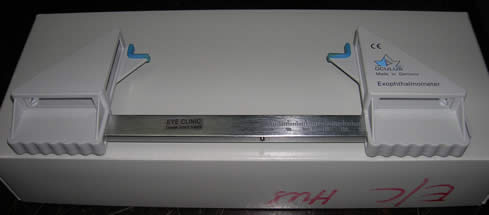
Infiltrative signs include: exophthalmos, conjunctival injection, chemosis, fullness of the eyelids, enlargement of lacrimal gland, herniation of orbital fat, infrequent blinking (Stellwag’s sign) increase intraocular pressure,35 strabismus and restriction of extraocular movements (Ballet sign). The inferior and medial rectus muscle is commonly involved in TAO.36 TAO is the commonest cause of axial proptosis in adults. It can be unilateral or bilateral. Proptosis is usually measured by Hertel exophthalmometer (Figure 3). It measures the distance between the lateral orbital rim and the anterior surface of the cornea. The upper limit of normal for whites is 18 mm and for blacks is 21 mm and for Asians the normal range is 12-18 mm. A difference in reading of more than 2 mm between the two eyes is suggestive of proptosis.
Figure 1. A patient with TED and corneal perforation of the left eye.
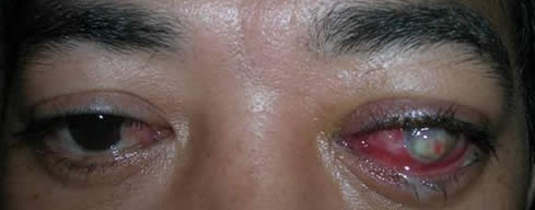
Figure 2. A patient with TED and strabismus of the left eye (esotropia).
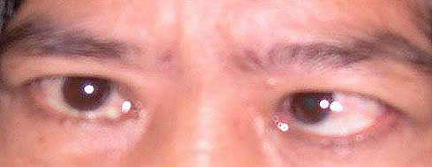
Figure 3. Hertel exophthalmometer.
THYROID DERMOPATHY / PRE-TIBIAL MYXEDEMA
It is also an autoimmune manifestation of Graves’ disease, which is usually associated with severe ophthalmopathy. Ophthalmopathy is said to occur first and dermopathy occurs later. Lesion of pre-tibial myxedema is usually asymptomatic and has only cosmetic importance. Thyroid-stimulating hormone receptor antibody in the connective tissue is believed to be the antigen initiating the immune process.37
COMPLICATIONS
- Inability to close the eyelids as result of lid retraction and proptosis can lead to exposure keratopathy.38 Corneal defects in TAO also occurs because of widening of the palpebral fissure accelerating evaporation of the tear film and increase in the tear film osmolarity.39
- Compression of the optic nerve or its blood supply due to enlarged extraocular muscles at the orbital apex causes optic neuropathy.40 Compressive optic neuropathy occurs in less than 5% of patients.41 The optic neuropathy produces progressive loss of visual acuity, decreased color vision and defects in the visual field, usually central scotoma.42
- The prevalence of glaucoma is said to be much higher in patients with Graves’ disease than in the general population.43
